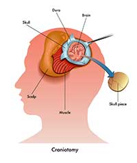
 During a craniotomy, a part of bone in the skull (known as a bone flap) is removed in order to expose the brain. This uses specialized tools and may involve a frame or markers placed on the scalp or intraoperative monitoring to reach exact locations in the brain. Scans may also be taken to provide images inside the brain. Once the brain is exposed, a number of procedures may be performed in situations such as:
- Removing blood clots
- Diagnosing or removing brain tumors
- Clipping or repairing an aneurysm
- Draining an brain abscess
- Removing an arteriovenous malformation or other brain lesion
- Repairing a fracture
- Relieving intracranial pressure
- Treating epilepsy
- Implanting devices to treat functional problems, like dystonia or Parkinson’s disease

The bone flap is replaced after the procedure is done and is held in place with sutures, wires, or plates. There are risks to a craniotomy, like with any other procedure, and vary according to the area of the brain that is being operated on. For example, speech may be affected if the affected area of the brain is responsible for speech.
There are some variations of a craniotomy, like a craniectomy (where a portion of the skull is permanently removed) and endoscopic craniotomy (which is performed with a lighted scope placed into a small incision).
A craniotomy generally requires a hospital stay, and the incision heals about three to four weeks after the procedure. It is important to follow the pre- and post-procedure instructions provided by your doctor.


